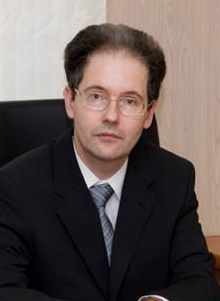
Head of the Department
Dmitrii E. Korzhevskiy, доктор медицинских наук, профессор РАН
e-mail: morphol@iemspb.ru
Phone: (812) 234-24-38
The Department consists of:
History of the Department
Nowadays, the Department of General and Particular Morphology is the only morphological subdivision of the Institute of Experimental Medicine (IEM).
The history of the Department had started from the Sector of Morphology and Pathomorphology of IEM, which had been organized in 1932 and had been consisted of 3 departments – the Department of Human Morphology (N.D. Bushmakin, Head), Department of General and Particular Morphology (A.A. Zavarzin, Head) and the Department of Pathomorphology (N.N. Anichkov, Head).
The Department of Pathological Anatomy was the first morphological department, which had been organized in the year of the Institute foundation (1890) and existed in the structure of IEM to the end of 80s of ХХ century.
The Department of General and Particular Morphology was founded by the academician A.A. Zavarzin in 1932. It was the center of development of theoretical histology in our country from the very beginning.
Famous concepts of tissue evolution: the theory of parallelism by A.A. Zavarzin and the theory of degenerative evolution by N.G.Khlopin had been developed in this Department. Professor D.N. Nasonov formulated the protein theory of insult and irritation of cells.
Structure of the Department had changed several times all over its’ history. As the result, the Department of Morphology existed as separate laboratories for some time. Different famous scientists had been working there, such as A.A. Braun, L.N. Ginkin, A.A. Zavarzin, Jr., G.S. Katinas, A.G. Knorre, V.P. Mikhailov, G.A. Nevmyvaka, P.G. Svetlov, S.I. Shchelkunov, G.V. Yasvoin, G.S. Strelin, etc.
The Department of Morphology was recreated from two independent laboratories of the Institute in 1980 (Corresponding Member of RAMS V.A. Otellin, Head).
The Laboratory of Experimental Histology (professor V.P. Mikhailov was the Head of the Laboratory) and the Laboratory of Cytology (which had been headed by Professor A.A. Manina) entered the Department. The main attention of the staff of the Laboratory had been paid to the studying the mechanisms of regenerative histogenesis and the functional morphology and neurotransmitter interactions in the central nervous system.
In 2007, the Department was reorganized by the academician of RAMS V.A. Nagornev and was named as The Department of General and Particular Morphology. From that time, the Department consisted of two laboratories: The Anichkov Laboratory of Atherosclerosis (academician of RAMS V.A. Nagornev was the Head of the Department) and the Laboratory of Functional Morphology of Central and Peripheral Nervous System (D.E. Korzhevskii, MD, Head).
The Anichkov Laboratory of Atherosclerosis originated from the Department of Pathological Anatomy. N.V. Uskov who had been one of the biggest pathologists in Russia became the first Head of the Department. Academician of AMS of USSR N.N. Anichkov had been headed the Department of Pathological Anatomy from 1920. He was the first to study the morpho- and pathogenesis of atherosclerosis. Professor M.V. Voyno-Yasenetsky had become the Head of the Department after the N.N. Anichkov death in 1964. He had been successfully and comprehensively studying the pathogenesis of acute infection diseases. Professor V.E. Pigarevskiy was the last Head of the Department of Pathological Anatomy from 1977 to 1989.
A number of studies crucial from the theoretical and practical points of view have been held in the Department during this period. They were devoted to non-specific resistance of human body to infections. In the course of these studies, previously unknown phenomenon of resorptive cellular resistance has been discovered and the concept of enzymatic and non-enzymatic cationic proteins has been formulated. The lisosomal and cationic test was created, which still has a great practical importance for prognostication and rapid assessment of the effectiveness of treatment of the range of the infectious diseases.
From 2010, P. V. Pigarevskiy became the Head of the Department of General and Particular Morphology.
Main Areas of Research
Nowadays there are two main directions of investigation:
- Studying of cellular-molecular mechanisms of the adaptive immune response during the atherogenesis and exploration of the immunopathological mechanisms of destabilization of atherosclerotic plaque.
- Studying of processes in the organs of nervous system after impact of different damaging factors.
In the framework of first direction, it was managed to develop and to substantiate the concept about the leading role of immunopathological factors in formation and developing of atherosclerotic damages of the artery of the human.
It was shown that deposition and formation of subendothelial layer of the intima if the arteria of modified forms of low-density lipoproteins, autogenic features, stimulating production of the endothelial cells of anti-inflammatory cytokines and adhesion molecules, connecting with ligands of monocytes and lymphocytes.
Activated monocytes and T-lymphocytes, penetrating into the intima in the very early stages of the atherogenesis with the help of cytokines launch the cascade of inflammatory reactions during which all the cells of the vascular wall are involved.
It is proved that developing of chronic immune inflammatory in the arteries during the atherogenesis in the human body is connected with the cellular reactions in regenerative lymphoid nodes.
Nowadays, the process of comparative studying of features of immune-inflammatory process in different atherosclerotic damages in human started with the aim of detecting of the reasons and mechanisms of destabilization of atherosclerotic plaques.
The aim of the Laboratory is to detect of predicates and clinical markers of destabilization that as the result it can allow. It can allow to at measures of predicting and treatment of high of coronary syndrome at the end. The integrated approach is used for the problem solving with simultaneous using of histological, immune-biochemical and immunimorphological and morphometric methods of the research.
The second line of research, which is carried out in the Laboratory of Functional Morphology of Central and Peripheral Nervous System, is aimed to study the structural organization and cytochemical features of nerve and glial cells of animals and human in the early ontogeny in pathology and aging by using immunohistochemical markers, fluorescent and confocal laser microscopy. Changes of the structural organization of organs of nervous system during the damaging influences and innervation pattern of vital organs of cardiovascular and digestion systems are also studied. Research of histogenesis and regeneration of the CNS and PNS is conducted on the model of transplantation of neural cell progenitors and mesenchimal stem cells into the damaged nerve.
Ontogenetic changes of the structural organization and functional state of glial elements of the brain, spinal cord, spinal ganglia and nerve structures of some internal organs (pancreas, heart, etc.) are studied. Our study revealed pattern of differentiation and change of glial cell phenotypes in ontogeny and their reactivity in the age aspect. Fundamental studies in this direction are crucial for understanding of the role of the glia in the pathological processes on different phases of the ontogeny, including its role in the developing of the neuroinflammation.
With the aim to integrate the topics of two laboratories of the Department, an investigation of arteria innervation during the developing of the atherosclerotic lesion of vessels, including brain vessels, is going to held, which should contribute to reveal the cell-molecular mechanisms of the development of stroke and diabetic macroangiopathy in the human.
Thus, as throughout its history, the main challenge of the Department of the General and Private Morphology remains unchanged: it is the solving of fundamental problems of experimental and compared morphology, as well as, studying of the pathogenesis of crucial social significant human diseases.
Key Academic Achievements
There close link was identified between the infiltration of vascular wall of the immune-inflammatory cells and the processes of the destabilization of the atherosclerotic plaque. It is established that in the zone of formation of the unstable atherosclerotic plaques there are serious dystrophic changes in tissue of the aorta wall which correlate with dystrophic changes of nervous apparatus and vasa sacorum.
These changes are coupled with this severe immune-inflammatory reaction, which is developing in the intima and in the adventitia of the vessel at the same time.
In the framework of research of morphofunctional changes in organs of nervous system in ontogeny and during damages, using the model of the transitory focal rat cerebral ischemia, we demonstrated for the first time a disorganization of astroglial component of the forebrain subventricular zone (SVZ), migration of microgliocytes through ependyma into the cavity of cerebral ventricle, and formation of atypical stellate smooth muscle cells in SVZ. Revealing of specific ependymocytes and neurons containing intranuclear beta-catenin allowed to hypothesize that the area of the medial habenula ependimal layer can be treated as the reserve neurogenic zone of diencephalon. By using the neural transplantation, we found that embryonic neocortical anlagen developing in an unusual biological niche demonstrated changes in cell cycle of the neuroepithelial cells, that correlates with impaired distribution of cadherin-immunoreactive cell contacts and the disorder of interkinetic migration of nuclei. Bone-marrow mesenchymal stem cells (MSC) injected into the brain were shown to have fibroblast-like growth during the first week of observation. After injection of MSC into the nerve, they remain viable for a week and then some of them differentiate into adipocytes and cells of perineurium. Using immunohistochemical methods for the detection of peripherin, PGP 9.5 protein, and tyrosine hydroxylase, new approaches have been developed for the selective detection of nerve apparatus in various internal organs of humans and laboratory animals.
Theses of the research assistants of the department over the past 5 years
Doctorate theses:
Candidate’s theses:
Academic titles received by the research assistants of the department over the past 5 years:
Patents obtained by the research assistants of the department over the past 5 years:
- Chumasov E.I. Korzhevskiy D.E., Petrova E.S. Immuno-histochemical way of identifying of sympathetic and parasympa-thetic structures in histological preparations. Patent of RF №2657787 from 28.12.2016. Published: 15.06.2018. Bulletin. № 17
- Antimonova O.I., Korzhevskiy D.E., Shavlovsky M.M. The way of fluorescent identification of amyloid. Patent №2673815 from 30.11.2017. Published: 30.11.2018. Bulletin. №34
Technologies obtained by the research assistants of the department over the past 5 years:
The most significant publications over the past 5 years
- Чумасов Е.И., Колос Е.А., Петрова Е.С., Коржевский Д.Э. Иммуногистохимия периферической нервной системы. СПб.: СпецЛит, 2020. 111 с.
- Пигаревский П.В., Мальцева С.В., Танянский Д.А., Фирова Э.М., Татаринов А.Е., Чивикова Н.В., Шовковый А.В., Мещерякова Н.Г., Федорова В.Ю. Матриксная металлопротеиназа 9 типа в крови больных ишемической болезнью сердца // Цитокины и воспаление. 2020. Т.19. №1-4. С.41-44.
- Abdurasulova I.N., Matsulevich A.V., Kirik O.V., Tarasova E.A. Ermolenko E.I., Korzevskii D.E., Klimenko V.M., Di Pierro F., Suvorov A.N. The protective effect of Enterococcus faecium L-3 in experimental allergic encephalomyelitis in rats is dose-dependent // Nutrafoods, 2019, 1:1–11. DOI 10.17470/NF-019-0001.
